GraeaeX
The premier solution for bringing the beauty and complexity of your 3D scan data to life.
GraeaeX helps transform scientific scan data into clear, textured visual outcomes for research, presentation, education, and physical display.

Interactive Data Interpretation
Compare related scan-derived views using the slider. Drag the control to examine structural differences between the source image and selected interpretation.
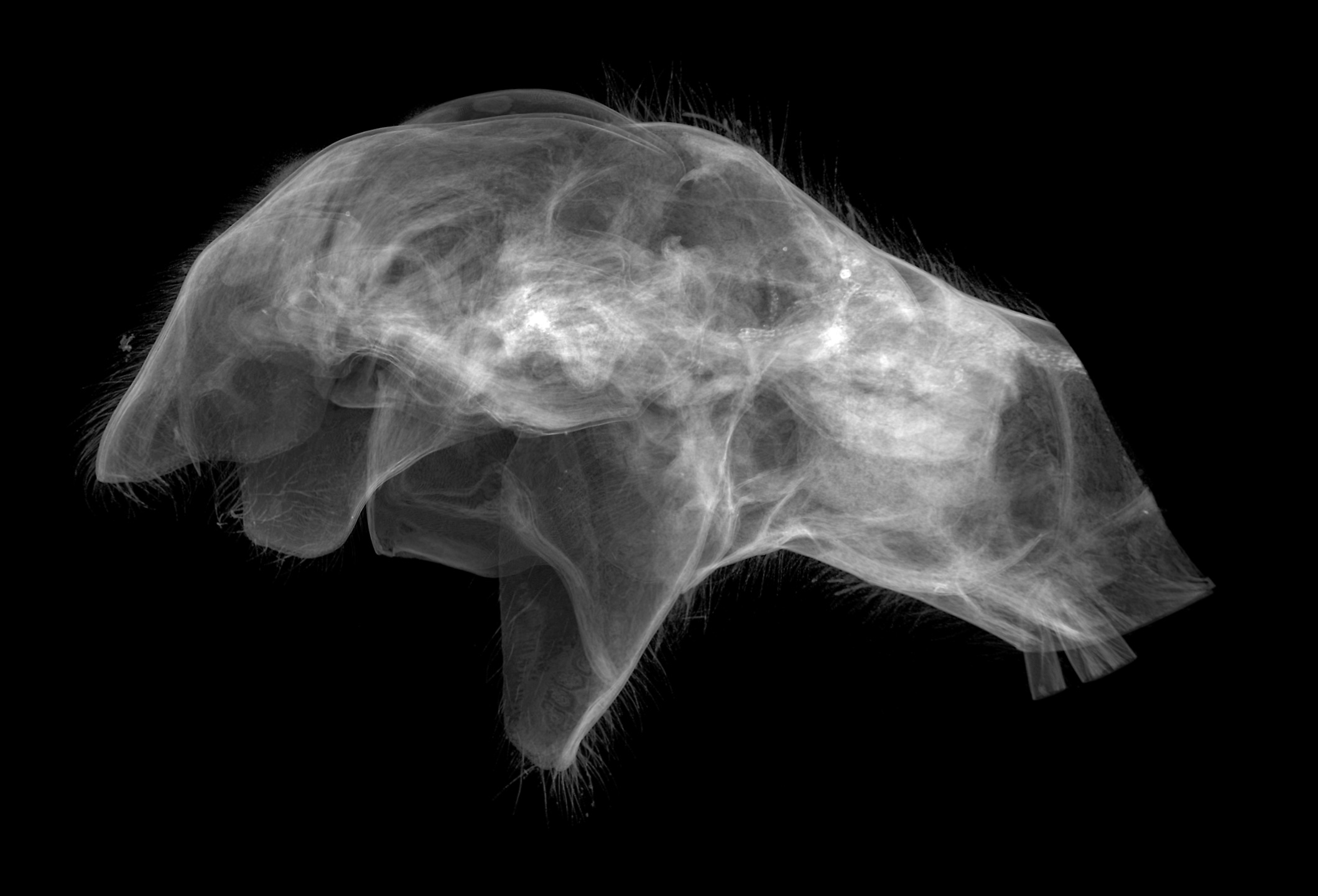 Base X-Ray View
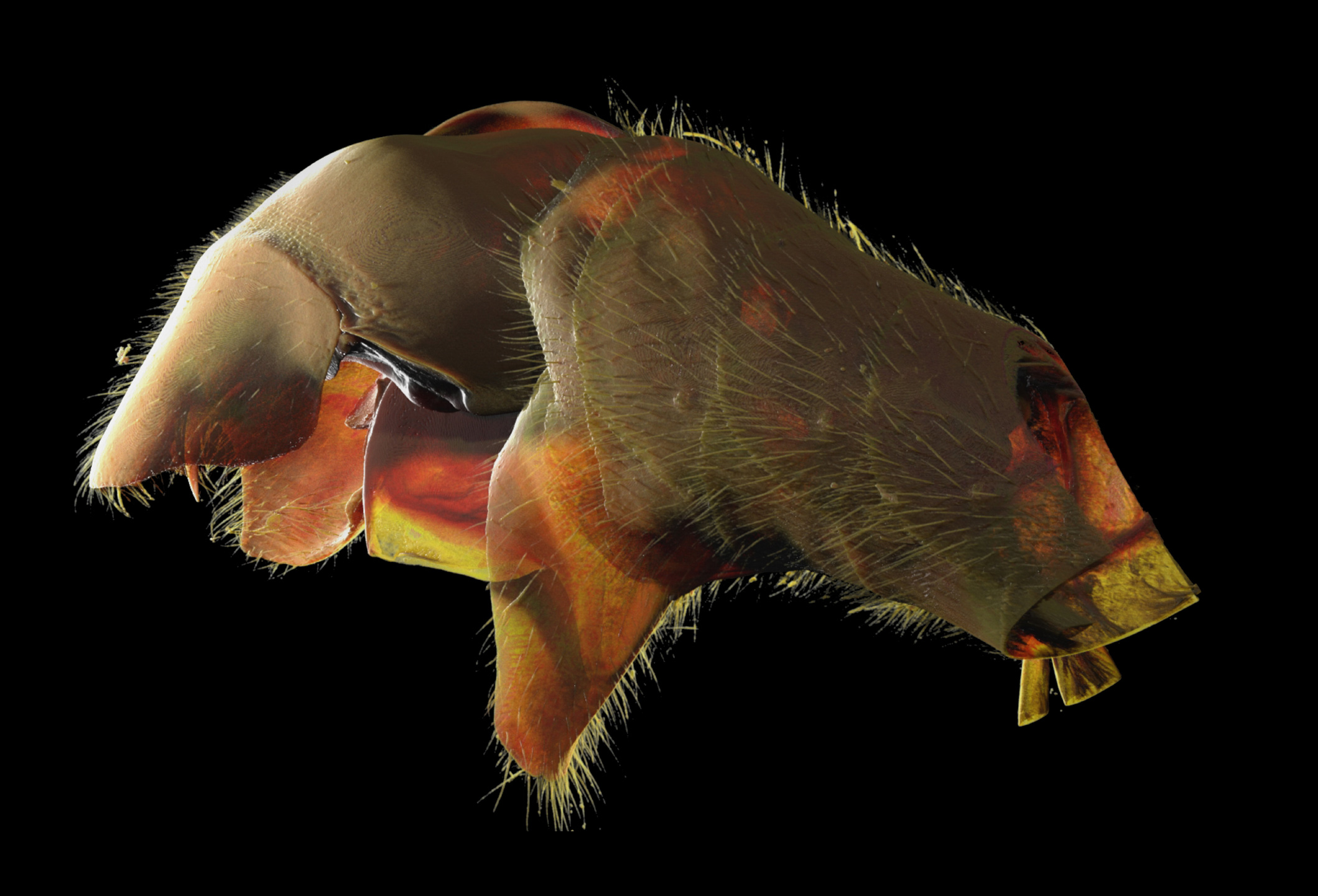 Density Map View
Specialized 3D Scan Processing
GraeaeX specializes in processing three dimensional scans into custom, fully textured two- or three-dimensional visual outcomes.
GraeaeX uses data generated by CT, PET, Sonogram, and related scan sources to produce realistic images, display models, and full color 3D prints.
Multi-Source Compatibility
Direct processing of CT, PET, Sonogram, and micro-CT data inputs.
High-Definition Texturing
Crafted color palettes and anatomical clarity for academic publishing.
Display & Presentation
Publication-ready render outputs for journal articles, museum exhibits, and lectures.
Physical 3D Prints
Full color physical 3D print models optimized for anatomical accuracy.
Approaching Realism – What you can do with 3D X-ray Microscopy?
TOSCA 2015, SESSION: CT – past, present and the future
Realism is a function of scale, resolution, material properties, and imaging capability. To approach realism—namely provide adequate visualization or representation for any material whether it be ant anatomy or 3D printed steel—requires having the imaging capability at the appropriate scale and spatial resolution. A new field of 3D X-ray microscopy (XRM) has emerged bringing dramatic resolution and contrast improvements to X-ray tomographic imaging. Analogous to computed tomography (CT), a specimen can be imaged without physical sectioning and a complete 3D view of the object is generated. Yet X-ray microscopes provide superior spatial resolution down to the nanoscale and tunable phase contrast to image materials such as nanoscale pores in filtration membranes or entire model organisms such as zebrafish ex vivo up to tens of centimeters in size. This is possible because laboratory-based X-ray sources have been coupled with high resolution optically coupled detectors and in some cases X-ray focusing optics to acquire tomographic datasets with resolution down to 50 nm across a great span of sample dimensions.
This talk highlights the imaging capability of a 3D XRM with application highlights from materials and life sciences. In the first example, tomographic data was collected by ZEISS Xradia 520 Versa of an ant specimen (Adetomyrma bressleri) for morphological studies to resolve species phylogeny. In this case, to ‘approach realism’ required intuitive and advanced visualization by GraeaeX. Secondly, for materials research, datasets collected at nanoscale resolution by Xradia 810 Ultra provided realistic microstructure input to improve computational transport models for polymer electrolyte fuel cells. Coupled with advanced visualization methods, three-dimensional X-ray microscopy (XRM) is a powerful sub-surface imaging technique that reveals tomography of microstructure from a range of materials, non-destructively.
Citations & References
- A. Merkle and J. Gelb, Microscopy Today 21, (2013) pp. 10-15.
- A. Tkachuk, et al., Z. Kristallogr. 222, (2007) pp. 650-655.
- S. Litster, et al., Fuel Cells 13, (2013) pp. 935–945.
Gallery of Scientific Visualizations
Select any visualization card below to view the high-definition scan render and detailed specimen metadata.
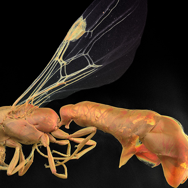 MICRO-CT PROFILE
Full Ant Profile
Adetomyrma bressleri Lateral Scan
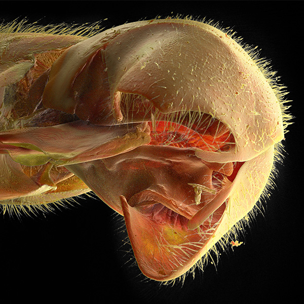 ABDOMINAL VIEW
Abdomen Underside
Ventral Abdominal Morphology (v09)
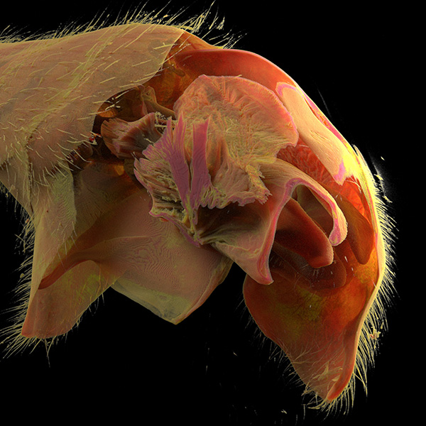 TERMINAL MORPHOLOGY
Telomere Structural Render
Terminal Segment Morphology
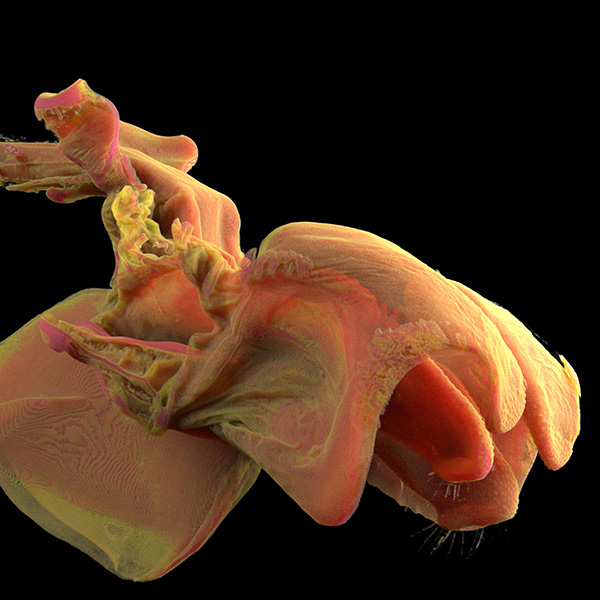 EXTENDED GENITALIA
Aedeagus Render
Male Extended Genitalia Scan
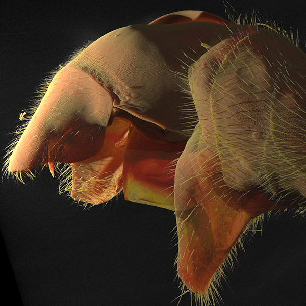 INTERNAL TISSUE 5K
Gonad 5K Render
Internal Tissue Micro-Structure
Selected Publications & Technical Reference
Approaching Realism – What you can do with 3D X-ray Microscopy?
TOSCA 2015: Tomography for Scientific & Industrial Applications, CT Session
Examines 3D X-ray microscopy (XRM) spatial resolutions down to 50 nm, combining Xradia 520 Versa tomography of Adetomyrma bressleri ant specimen morphology with GraeaeX 3D visualization.
ZEISS Atlas 5: Multi-Scale Correlative Workspace
Multi-modal SEM, FIB-SEM & XRM Integrated Workflow Engine
Overview of 3D nanotomography, automated array tomography, xROI definition, and multi-scale image correlation across light, electron, and X-ray imaging sources.
ZEISS Xradia 520 Versa 3D X-Ray Microscope
Sub-micron Non-Destructive 3D Imaging System
Details true 0.7 μm spatial resolution, 70 nm voxel size, Resolution at a Distance (RaaD) optical magnification, and DSCoVer dual-scan energy visualizer.
Contact
For project inquiries, collaborations, archival questions, or scientific visualization work, contact GraeaeX.